EYE EXAMINATIONS
We recommend having an eye examination at least every two years, or more frequently if advised by one of our optometrists.
The importance of regular eye tests is not solely to determine the need for glasses, but also, to assess the general health of the eyes. Signs of many underlying health conditions such as diabetes, high blood pressure, and high cholesterol can be detected during the eye examination.
At Mullins & Henry we believe that no two pairs of eyes are the same, and so we adjust our eye examinations to meet your individual needs. We don't rush our eye tests and make sure our appointments include enough time for you to ask us any questions you may have.
WHAT HAPPENS IN AN EYE TEST?
During your eye examination, your optometrist will assess your vision to determine if you require spectacles, and, if so, determine the correct prescription. The examination will also involve checking to see if your eyes are healthy.
Our optometrist will begin your eye examination by asking you a few questions. This will include any problems you may be experiencing, e.g. headaches, or difficulty seeing up close or in the distance.
Your appointment will also include a discussion on your general health and medical history. It is important that we are aware of this as it can have a direct effect on the eye and vision, for example:
-
arthritis may cause dry eyes or other problems (i.e. difficulty in reading)
-
someone on long-term steroid treatment may have raised intraocular pressure (i.e. glaucoma)
-
family history is also important. Certain conditions may be hereditary such as glaucoma and diabetes.
OUR EYE TEST TECHNOLOGY
At Mullins & Henry, we pride ourselves on ensuring every patient experiences a meticulous eye examination. We use various pieces of optical equipment to test different aspects of your vision and the health of your eyes.
Below we explain what the equipment is and what they do. Each one of these auxiliary tests may not necessarily be carried out on every patient. Your optometrist will decide during your appointment which ones are necessary.

RETINOSCOPY
This is a method the optometrist uses to determine the prescription you may need without actually getting you to read the letter chart, i.e. it is an objective test.
The optometrist uses a retinoscope to shine a light on the back of the eye (the retina) and he/she sees a red reflection called the retinal reflex. When the optometrist moves the retinoscope this reflex moves also. He/she will put test or trial lenses in the trial frame in front of the eye until the reflex stops moving. This is called neutralisation and gives the objective result of the power of the spectacles needed for the patient to see clearly.
This is a very helpful procedure where the patient may be unable to communicate to the optometrist, e.g. a mute patient or a very young child.
Some optometrists may use an auto-refractor to determine this objective result. This is an automated instrument, which will give an estimation of the prescription required, it is useful in giving the optometrist a starting point for his/her subjective test.
SUBJECTIVE TEST
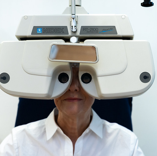
The optometrist will determine the final strength of the spectacles required. In this case, you are fully involved (i.e. it is subjective) and will read the letters on the letter chart for the optometrist.
The optometrist will show stronger and weaker lenses to you until the best vision is obtained. The optometrist will also determine if there is any astigmatism present by using a lens called a cross/cylinder.
The final result is fine tuned by using a duochrome, or red/green test. Each eye is tested separately first and the final prescription is determined binocularly, i.e. with both eyes together.
OPTHALMOSCOPY
The optometrist uses the ophthalmoscope to view inside the eye. This instrument has a series of tiny lenses on a revolving rack and by changing these the optometrist can focus on the front of the eye (the cornea) right through to the back (the retina). This part of the examination is a vital health check for the eye. It can reveal conditions such as cataracts, glaucoma and retinal detachment.
The eye is the only part of the body where the blood vessels can be seen without surgery. The optometrist can see the arteries and veins in the retina and may therefore be able to detect signs of vascular problems such as high blood pressure and diabetes. However, the optometrist is not qualified to diagnose these problems so if he/she detects anything abnormal he/she will refer the patient on to their G.P. or directly to a specialist if necessary
SLIT LAMP EXAM
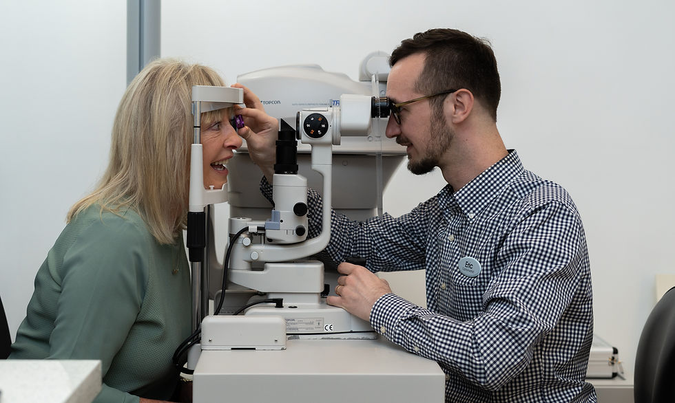
This is a large table mounted instrument and is used to view the cornea and eyelids and anterior part of the eye in more detail than the ophthalmoscope. It is a binocular microscope with variable magnification and light source. It is used most frequently in contact lens fitting but it is also very useful as part of the general eye examination. The optometrist may also use this in conjunction with a small but powerful lens called a Volk lens to look at the retina.
VISUAL FIELD TEST
This test is used to test the central and peripheral fields of vision and to determine if there are any “gaps” in this field of view. If the field of vision is not full or complete it could indicate problems such as detached retina or a hemorrhage caused by high blood pressure. Glaucoma will also affect the visual fields, as can many neurological conditions.

RETINAL IMAGERY
Retinal imaging is the process of taking a digital image of the back of your eye. It depicts the retina (where light and images are reflected), the optic disc (a region on the retina that houses the optic nerve that transmits information to the brain), and blood vessels.
Our optometrists can receive a considerably larger digital view of the retina through retinal imaging, using a piece of technology called a Fundus Camera.
This camera enables our optometrist to take a high-resolution digital photograph of the back of the eye. The resulting image enables you to see what the optometrist can see when they look in the back of your eyes. Most importantly, a permanent photographic record of your eye health is created, which can be reviewed and compared at further eye examinations to monitor even the smallest change that can indicate an issue.